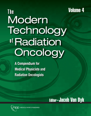
The Modern Technology of Radiation Oncology, Vol 4
Author: Jacob Van Dyk, EditorISBN: 9781951134020
Published: 2020 September | 522 | Hardcover
Price: $ 175.00
Physical and Engineering Sciences in Medicine | June 2021
Medical Physics Publishing recently published a book entitled “The Modern Technology of Radiation Oncology”, edited by Jacob Van Dyk this is the fourth volume in a series of textbooks which informs our professional community as we embrace the Technology Evolution Revolution in the Field of Radiation Oncology.
Medical Physics Publishing is not for profit publisher that was founded by the medical physics visionary Professor John Cameron in Madison Wisconsin in 1985. In my opinion they continue to provide unparalleled quality textbooks that support our medical physics profession.
Technology is evolving so rapidly that each textbook volume informs us of new essential current and topical advances in the field. In my opinion, this volume along with its predecessor is a must-read for all professionals to stay informed about current trends in technology being introduced into the radiotherapy clinic.
The Textbook consists of eighteen invited chapters from contributing authors that are experts in the field. Given below is a list of chapter titles and a very brief comment on content within each respective chapter.
Chapter 1 “Technology Evolution in Radiation Oncology: the Rapid Pace Continues”. Is written by Jacob Van Dyk. This chapter nicely sets the scene for future chapters. For example trends in innovations related to hybrid imaging and treatment technology. New innovations such as Radiomics and Artificial Intelligence are also introduced. Figure 1.1 has a particularly strong impact as it graphs clinical benefit against time from 1895 to 2020 and includes images of various technology innovations.
Chapter 2 “Surface Guidance in Radiation Therapy”. This chapter outlines the stereoscopic surface guidance technology designed to track patient surface contours in real time. This chapter includes clinical implementations, clinical evidence, quality assurance and end to end testing of these systems. This chapter is a prime example of a concise practical reading resource for a relatively newly implemented clinical technology which other medical physicists may be currently implementing in their clinic.
Chapter 3 “PET/MRI as a tool in Radiation Oncology”. Gives an outline of the hybrid technology and summarises its potential in treatment response monitoring. This chapter gives an excellent perspective of the use of this technology in breast, brain, prostate and liver.
Chapter 4 “Real-Time Image Guidance with Magnetic Resonance”. Outlines the new clinically deployed hybrid MRI-linac Radiotherapy machines that offer a new image guidance paradigm. The chapter includes a summary of the various system designs along with adaptive radiotherapy treatment strategies employed. The potential to apply MR imaging for response monitoring is discussed. Dose distributions are effected by the B field Lorenz force, and some bespoke quality assurance and dosimetry is required for these machines. As this is an area of my current research I have to say this chapter was my favourite one.
Chapter 5 “Stereotactic Body Radiotherapy”. Gives a review of the history and lineage of stereotaxic and stereotactic therapy! The transition from frame to frameless systems is discussed. Flattening filter-free Linear accelerators, CyberKnife, Gamma Knife and Zap-X are discussed. Clinical MRI-linac systems are also discussed. Dosimetry required for small fields is reviewed along with clinical sites to which SBRT is most applicable.
Chapter 6 “Radiation Uncertainties: Robust Evaluation and Optimization”. Covers margin recipes and methods to evaluate dose uncertainties along with optimization methods for robust treatment planning.
Chapter 7 “Automated Treatment Planning”. Outlines how automation improves efficiency, quality and safety. Figure 7.2 compares a conventional planning work flow with an automated planning workflow which is simpler when deployed. The progress with automated contouring and quality assurance checks for automated planning are discussed as are current clinical implementations of automated treatment planning systems.
Chapter 8 “Artificial Intelligence in Radiation Oncology”. Gives a brief history and overview of Deep Learning and gives indications where this can be applied to Radiation Oncology based problems. Examples include Auto-segmentation of targets and organs at risk, applications in knowledge- based planning, and potential applications to Radiomics analysis. There are also sections on the role of AI as a new tool in clinical trials and data-driven outcome modelling.
Chapter 9 “Adaptive Radiation Therapy (ART)”. Covers offline ART and online ART including state-of-the-art tumour tracking protocols. Clinical trials for clinical sites that have deployed adaptive strategies for various treatment sites are also summarised. Practical considerations about workflow staff and uncertainties and limitations of ART are also discussed. This chapter is written by the Australian Image X Institute group that are world leaders in motion adaptive radiotherapy.
Chapter 10 “Machine Learning in Radiation Oncology: What have we learned so far”. This chapter complements chapter 8 with more applied examples. Comparative examples of contouring head and neck patients using machine learning compared with Oncologist defined contours are reviewed. Regulatory requirements for machine learning algorithms for performance and clinical evaluation are also discussed.
Chapter 11 “Applications of Big Data in Radiation Oncology”. As they point out is “one of the most hyped terms in recent times” and the chapter defines the features of big data: Velocity, Variety and Veracity. The use of Big Data for better outcomes in decision support and precision medicine is outlined.
Chapter 12 “Quantitative Radiomics in Radiation Oncology”. This is a detailed comprehensive review of the current state of the Art of Radiomics. Radiomics applied to CT, PET, and MRI data sets are reviewed as are practical considerations such as standards, reporting, and safeguards.
Chapter 13 “Radiobiological Updates in Particle Therapy”. The chapter presents the laboratory and experimental evidence for relative biological effectiveness (RBE) and then covers its clinical application in Proton and Heavy Ion (e.g. Carbon-12) Radiation Therapy. The chapter then outlines evidence from proton treatments and future clinical use of RBE for biological optimization.
Chapter 14 “Radiation Oncology using Nanoparticles with High Atomic Numbers”. Gives a summary of Nanoparticle design, delivery considerations, and dose enhancement factors when combined with Radiation Therapy for dose enhancement.
Chapter 15 “Financial and Economic Considerations in Radiation Oncology”. Outlines ways to quantify the cost of Radiation Oncology using models that equate economic costs with improved outcomes. Various Cost-Effective Analysis (CEA) methods are presented such as the Quality Affected Health Year QALY. One QALY equates to one year of perfect health, I can’t remember one of those recently. This chapter links nicely with chapters 16 and 17 by introducing the frameworks for analysis.
Chapter 16 “Global Considerations for the Practice of Medical Physics in Radiation Oncology”. This chapter outlines professional and academic aspects of medical physics and frames the evolution of medical physics education and professional training. It importantly outlines the Global medical Physics Issues and the disparities that exist while outlining initiatives that are attempting to address these issues.
Chapter 17 “Emerging Technologies for Improving Access to Radiation Therapy”. This chapter highlights that only about 10% of the global population has access to Radiation Therapy resources! The chapter summarises situational radiotherapy resources by Country and Globally. Barriers such as economic and technological resources along with minimal requirements to sustain a radiotherapy service are discussed. The chapter concludes with some novel emerging technologies in radiotherapy that may aid in the democratisation of radiotherapy services on a Global scale.
Chapter 18 “FLASH Radiation Therapy: A new Paradigm”. It is fitting the last chapter of the text book outlines the potential use of ultra-high dose rate radiotherapy (FLASH). The preclinical evidence using FLASH points to an improved Therapeutic Ratio with potential reduction in normal tissue toxicity. This chapter outlines the evidence and potential dose rate thresholds and hypothesis for mechanisms related to FLASH.
Thank goodness medical physics has Jacob Van Dyk, like Tiger Woods and Phil Mickelson in golf his text books continue to make major comebacks. He has managed to assemble the most talented among us to sustain the up-todate knowledge that is essential to our profession. Reference knowledge from this text book will help ensure the medical physics profession is at the cutting edge of cancer research and clinical treatment.
This Text Book has taken pride of place on my book-shelf right next to my most treasured Porsche Magazines. I could not give it a higher accolade than that.
Peter E. Metcalfe
School of Physics and Centre for Medical Radiation Physics, University of Wollongong, Wollongong, NSW 2522, Australia
Physical and Engineering Sciences in Medicine
https://doi.org/10.1007/s13246-021-01027-w


