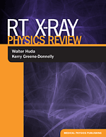
RT X-Ray Physics Review
Author: Walter Huda and Kerry Greene-Donnelly, AuthorsISBN: 9781930524545
Published: 2011 | 460 pp | Softcover
OUT OF PRINT
The Radiation Safety Journal – Official Journal of the Health Physics Society | Mar 2013
RT X-Ray Physics Review is an extensive (almost 500 page) volume that covers the x-ray physics component of the radiographic technologist (RT) certification examination given by the American Registry of Radiologic Technologists (ARRT). The authors point out that physics questions constitute approximately 60% of the written exam and are considered its most difficult segment. As a preparatory aid, each of the 15 chapters in the Review has its own multiple-choice exam of 30 questions. At the end of the text, there are two additional 100-question exams covering the entire topic of x-ray radiological physics. Each question has an answer supplied; usually there is a specific page reference to one (or both) of two standard textbooks in the RT field (SC Bushong, Radiologic Science for Technologists, 9th Ed. and RR Carhon and AM Adler: Principles of Radiographic Imaging, 4th Ed.). Thus, if a student gives an incorrect response to any question, they may generally investigate the relevant concept more thoroughly via the two references. Some five Appendices are also included in the Review. These tabulations cover physical and radiological units as well as scientific prefixes and their definitions. A final Appendix lists radiological physics, health physics, and other important medical websites.
Three major partitions of the Review occur: I) Basic Physics, II) Imaging with X-rays, and III) Dose, Quality and Safety. Part I (five chapters) includes concepts such as matter-energy, electrostatics, x-ray production, and interaction. Description of x-ray tubes and the inverse-square law are also covered. Magnetism is discussed but not used, as this is a strictly x-ray review. In Part II, the authors introduce detection of x-rays and how images (both analog and digital) are formed. Film and solid-state detectors are described. Included in this part are separate chapters on fluoroscopy and CT scanning. The latter is particularly good, with a clear description of the meaning of window and level leading into a discussion of Image GentlyTM and reduction of pediatric CT dose values. Part III continues this theme with a description of radiation dose, image quality and quality control, as well as radiation biology and radiation protection. The authors include air Kerma but also use Roentgen in their exposition. CTDI is defined, and its measurement is shown. Of these last five chapters, Quality Control contains a particularly strong discussion with specific illustrations of QC in mammography and CT scanning. The authors explicitly refer to requirements for accreditation by the ACR (American College of Radiology) in both these areas.
The Review is very well written with essentially no typographical errors. Figures and tables are generally clear with the sole exception of low-contrast CT resolution in Chapter 12. There are several errors of fact that any instructor can easily point out. Among these are unnecessarily rough formulae to convert between Celsius and Fahrenheit temperatures. The authors also confuse the X and Y chromosome. In several places in the Review, terms could have been defined more explicitly; e.g., free radical, stochastic, and voxel. This criticism, however, is not a strong one as the interested reader may use the two reference texts mentioned above and/or the internet to supply the desired background information.
Although the Review is well structured and covers all important topics thoroughly, several additional features and changes should be considered for later editions. Adding an index would probably be the single most important of these. Presently, the reader must hunt for topics (such as the half-value layer) using the Contents section. Certain questions, which presently have no references in the two texts listed, should be given a separate reference (from the current Bibliography) or have expanded discussion in any future revision. It is obvious that the current two referenced texts may eventually undergo revision, thus making cross-checking of topics more difficult. I would also recommend that discussion of Luminance and Illuminance be improved in the body of the future edition by inserting material presently sequestered in Appendix D. Finally, all question sets would be improved, I believe, if there were some arithmetic problems in the Review using hand calculators; e.g., deriving results from the exp(-µx) function. These would give x-ray students more confidence in their ability to determine numerical answers and not just in picking one verbal response from another in multiple-choice questions.
The Review serves its purpose well and would be a valuable aid in passing the x-ray physics segment of the ARRT examination. Its strong suit is the 15 sets of chapter questions (450 total) along with the two 100-question practice exams. By reviewing answers and reading specific pages in the two reference texts, the x-ray student is extensively supported in any potential self-study program. By going over chapter tests sequentially, one could find areas of weakness that would be remedied. However, use of this document along with pedagogic support by the student’s physics teachers is probably the ideal way to prepare for the AART examination. The Review could then be individually supplemented as needed with outside study materials, lab exercises, and handouts that describe any difficult concepts in alternative ways. Medical or health physicists are probably the requisite teachers of such x-ray students. Thus, the physics community should be aware of this Review and its obvious pedagogic advantages for numerical evaluation in the RT classroom. Authors Huda and Greene-Donnelly have performed an important task and deserve our thanks.
Lawrence E. Williams
Radiology Division
City of Hope National Medical Center


